- Press Office
- Eating egoistically
Eating egoistically
Marine algae produce large amounts of so-called polysaccharides, which form an important source of carbon for microorganisms. Typically, these long-chain sugar molecules are broken into smaller pieces before bacteria can take them up. However, that is not the only option, as Greta Reintjes from the Max Planck Institute for Marine Microbiology in Bremen now shows in a new publication.
On a research cruise to the Atlantic, Reintjes discovered that many marine bacteria use long-chain sugar molecules with a so-called “selfish” strategy. They first take up the large molecules and subsequently process them in a discrete section, breaking them into short-chain sugars. „The discovery of a widespread alternative substrate utilisation mechanism significantly affects our global estimates of carbon turnover by marine bacteria”, says Reintjes.
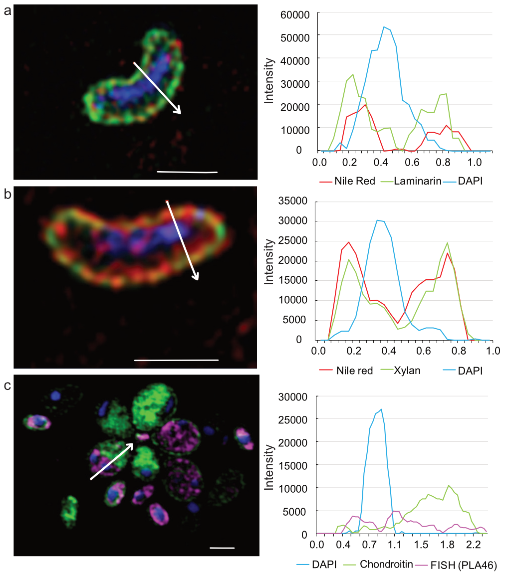
„With a highly efficient protein machinery, these marine bacteria take up short and long sugar chains through their outer membrane into the so-called periplasma”, explains co-author and MPI-director Rudolf Amann. The periplasma is situated between the outer and the inner membrane and is thus protected from food competitors. Using high-resolution light microscopy, Reintjes and her colleagues managed to depict fluorescently labelled sugar chains inside of the bacterial cells. „We discovered that only there the individual small molecules were further processed”, continues Amann.
Using molecular techniques, Reintjes identified the selfish bacteria as belonging to the Bacteroidetes, Planctomycetes und Gammaproteobacteria. „This mechanism was previously known from gut bacteria of the genus Bacteroidetes, but it has so far not been found in the marine realm”, says Amann.
The study now published in ISME Journal is part of Greta Reintjes Ph.D. Thesis, which she has defended at the MPI Bremen on April 28.
Original publication
Greta Reintjes, Carol Arnosti, Bernhard M Fuchs und Rudolf Amann (2017): An alternative polysaccharide uptake mechanism of marine bacteria.
The ISME Journal advance online publication, 21 March 2017; doi:10.1038/ismej.2017.26

Contact
Project leader
Department of Molecular Ecology
MPI for Marine Microbiology
Celsiusstr. 1
D-28359 Bremen
Germany
|
Room: |
2222 |
|
Phone: |

Head of Press & Communications
MPI for Marine Microbiology
Celsiusstr. 1
D-28359 Bremen
Germany
|
Room: |
1345 |
|
Phone: |
