- Departments
- Microsensor Group
- Projects
Projects
The microsensor group studies microbial ecology in a wide range of environments: seep systems in the deep-sea, coastal sediments, coral reefs, anoxic lakes, microbial mats, and animal associated microbes. The research is highly diverse, including photosynthesis, nitrogen and sulfur cycling, calcification, habitat mapping and cell physiology. Most themes involve the study of the functioning of intact microbial communities with non-invasive methods that allow as direct an observation as possible. Together with the MPI workshops, we continuously strive to develop novel methods, and apply them in our research. We collaborate widely with other institutes and with groups within the institute to share knowledge and ideas.
Reactive Oxygen Species
Olivia Metcalf, Dirk de Beer
Reactive oxygen species are fascinating compounds both essential and threatening to life. They consist of a group of small oxygen containing compounds which are intermediates in the reduction of oxygen to water and the reverse process, the oxidation of water to oxygen. They have a broad range of activity levels, ranging between the hyper reactive hydroxyl radical, which reacts with organic compounds within the cell at diffusion-limited rates, to the more selective superoxide radical, which focuses its attention on iron and iron-sulfur clusters within protein. In all cases, they can cause devastating damage to cells. However, emerging evidence also indicates that they perform essential functions within cells and organisms. For example, the growth of some heterotrophic bacteria was recently shown to be dependent on external superoxide by Hansel and colleagues in 2019. Superoxide has also been found to enable iron acquisition in some cyanobacteria and raphidophytes (Rose et al. 2005; Roe and Barbeau 2014; Garg et al. 2007).

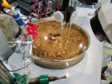
Olivia Metcalf is investigating the distribution and dynamics of reactive oxygen species within the keystone nitrogen-fixing cyanobacterium Trichodesmium and intertidal sediments of the Wadden Sea.
Transport and mineralization processes in intertidal zones
Beaches and plates
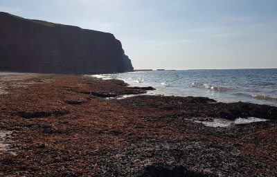
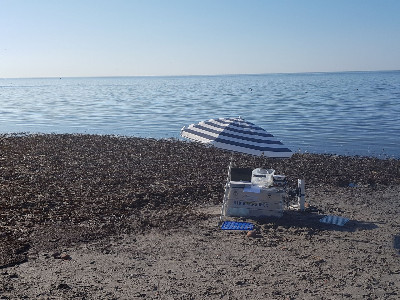
Kelp can accumulate in enormous amounts on beaches. This sudden input of organic material leads to a different degradation scenario as in normal early diagenesis. Marit has worked with Dimitri Meyer (Univ. Vienna) on a combined functional and biodiversity study. The intensity of the degradation rates allowed to detect all steps: hydrolysis, fermentation, aerobic and anaerobic respiration. Normally, degradation follows a series of thightly coupled steps, where products of anaerobic steps are oxidized by oxidized intermediates and eventually by oxygen. Whereas normally all stays internal in the sediments, from kelp deposits large amounts of reduced compounds escape, both organic and inorganic and partially toxic. For example, the sea adjacent to the kelp deposits become sulfidic, and massive amounts of sulfuroxidising mats develop quickly. This aerobic community was surprisingly diverse, and contained known anaerobes that adopted an aerobic lifestyle and had lost their denitrifying genes. The varying O2 levels induced by tides lead to significant CO development by reaction with the humic acids in the kelp deposits. CO is converted anaerobically via H2 by sulfate reduction and also is released to kill passing animals and tourists (www.bbc.co.uk/news/world-europe-14324094). The very sudden and strong increase in degradation processes upon kelp additions to sediment indicate the community is tuned to kelp and hungrily waiting for it. To demonstrate the specificity of the community, Marit is now studying the hydrolysis of kelp to specific sugars and the responses to different carbohydrates.
Sulfur bacteria
Charles Schutte, Verena Salman-Carvalho, Barbara MacGregor, Marit van Erk, Dirk de Beer
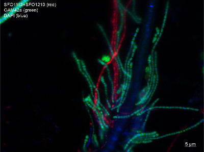
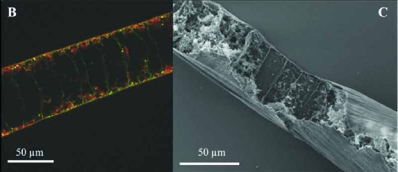
The sulfide oxidising Beggiatoa (nowadays called Beggiatoacea) are among the largest bacteria found. They form multicellular filaments with a length of cms and a thickness of upto half a mm. Most of their cell volume is a vacuole filled with upto 500 mM nitrate. Beggiatoa form large white mats on sulfidic and organic rich sediments. The filaments are highly motile, and migrate between the sediment surface and the sulfidic zone deep in the sediments. They survive the anoxia using the stored nitrate, which allows them to stay for a month in the anoxic zones.
We investigate how they orient themselves in the sediments (if you move you must navigate, e.g. to find the surface back), their phylogeny, how they oxidise sulfide, and how they grow.
An old debate is if Beggiatoa oxidises sulfide via denitrification or via dissimilatory nitrate reduction to ammonium (DNRA). Important as ammonium remains as nutrient in the ecosystem, nitrogen not. It appears they can do both.
Beggiatoa is in deep-sea vents the main primary producer of biomass. CO2 fixation is performed by RuBisCo. We showed that their growth rate is proportional to the CO2 partial pressure. That is much higher at 2 mm depth than at the surface. Thus migrating downwards supplies Beggiatoa with sulfide and CO2.
Phototrophic microbial mats
Dimitri Meyer (University Vienna), Arjun Chennu, Andreas Greve, Dirk de Beer
Microbial mats inhabited the world before the great oxidation event (GOE), and formed the oldest known fossils. Part of these mats were structured by Cyanobacteria, the microbes that can perform anoxygenic photosynthesis and 'invented' oxygenic photosynthesis. After the GOE, complex life developed that destroyed the mats by grazing. Nowadays, mats are only found at places where no higher lifeforms can survive: hot-, hypersaline or anoxic habitats. Anoxic illuminated habitats are very rare, as light leads to oxygen production, and are limited to sulfidic outlets of the deep biosphere. For more on this see www.mpi-bremen.de/Judith-Klatt.page.
Although the modern mats no doubt differ from those that made the 3 gA old fossils, we can learn a lot about the old world from them.
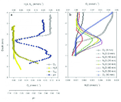
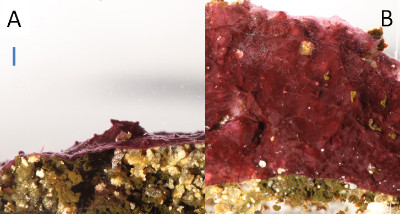
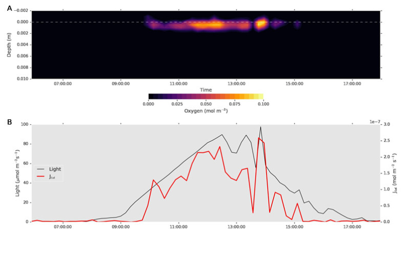
A highly interesting site is the 'Little Salt Spring' in Florida. This oasis of Florida's recent past natural beauty is currently surrounded by retirement residences, hospitals and funeral homes. Hopefully it can resist the developers. It is a sinkhole, funnel shaped and stratified. Below 2 m depth is perfect anoxia, and at the lake floor at 9 m depth red microbial mats grow inhabited by Cyanobacteria. We found that in this perfect anoxic habitat, a few hours per day oxygen developed. This sliver of oxygen in the upper mm of the mats can sustain the presence of aerobic bacteria (sulfide oxidisers and nitrifiers). Probably, this minute and ephemeral oxygenation in the upper layer of mats occured long before the GOE, and critically, prepared life for age of oxygen. Both defenses against reactive oxygen (e.g. catalase) and respiration on oxygen were ready by small populations that developed in these tiny pockets.
de Beer, D., et al., Oxygenic and anoxygenic photosynthesis in a microbial mat from an anoxic and sulfidic spring. Environ. Microbiol., 2017. 19(3): p. 1251–1265
Sabkha
Raeid Abed ([Bitte aktivieren Sie Javascript]), Andreas Greve, Arjun Chennu, Dimitri Meyer ([Bitte aktivieren Sie Javascript]), Dirk de Beer
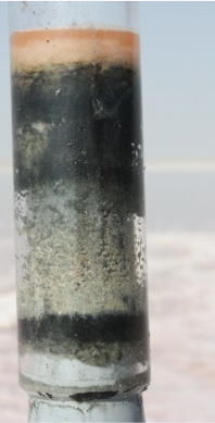
Sabkhas consist of salt-covered sediments, found in hot and dry climates. The salinity is saturated halite, yet they contain a diverse and dense microbiome. It was thought that the salt layer protects the deeper layers from dehydration and irradiation (Baas Becking, L.G.M., Geobiologie of inleiding tot de milieukunde. 1934, Den Haag: Van Stockum & Zoon; McKay, C.P., et al., PLoS ONE, 2016. 11(3): p. e0150342-e0150342).
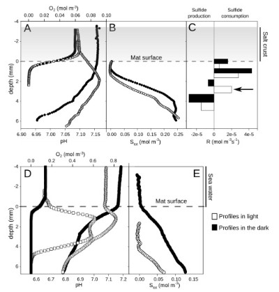
This appeared not true: radiation and gasses pass the salt unhindered.
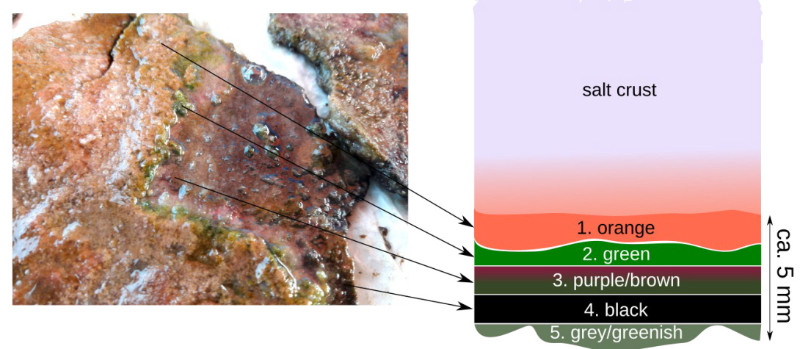
The microbes under the salt layer appeared not active. Only after inundation by seawater (as irregularly happens), high metabolic rates were observed (photosynthesis, respiration, sulfate reduction).
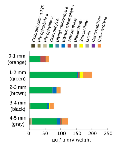
The two zones are observed in pigment distribution and light spectra, and molecular data show two different communities. The lower one is probably sulfide resistent, the upper one not.
Crucial is that the sabkha is hardly active during the dry period. No net primary production is possible, as anoxygenic phototrophy is coupled to sulfate reduction.
To sustain an active ecosystem periods of decreased salinity are needed.
Deep-sea studies LOOME, SUMSUN, MUMM, Guaymas
de Beer, Lichtschlag
All these deep-sea studies are joined Habitat-Microsensor group collaborations. The LOOME project (Wenzhöfer, de Beer) is a demonstration mission partially funded by the EU ESONET network that aims to lay the groundwork of deep-sea cabled networks. We test a variety of sensors placed on an observatory that can be coupled to a network. We want to study the phenomena before, during and after an outburst of a cold seep, thus sensors 'look' downward for early signs of an eruption (seismics, deep T sensors), at the sediment surface for enhanced porewater flow and effects on fauna (T-strings, various chemosensors, a time-lapse camera), and in the water column (CTDs, a scanning sonar). LOOME was deployed in July 2009 and will be recovered in September 2010. During the deployment of LOOME we tested the hypothesis that bioluminescence is elevated near seeps, as here primary production by chemosynthesis is high. Heinz Filthuth and Paul Färber built a supersensitive deep-sea photoncounter, that was deployed from a winch. The hypothesis had to be rejected, actually bioluminescence was reduced in the volcano area.
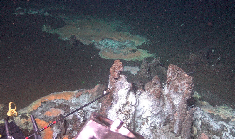
In the SUMSUN cruise (Boetius, de Beer) we investigated a natural CO2 seep, to assess the effect of intense CO2 seepage on the biogeochemistry, microbiology and fauna near the seeps. Preliminary data show a very low pH in the sediments, low microbial activities and absence of fauna near the seeps. The first in situ measurements ever done in such a habitat, showed that the sediments totally change upon retrieval due to CO2 outgassing, underlining the need for our in situ technology. Anna Lichtschlag (MUMM) finished her PhD studies on cold seeps that focused on the 'competetion' between biological and chemical sulfide oxidation. She has collected a treasure of data in the Nile fan, the Haakon Mosby and other seeps on the Norwegian shelf, and seeps in the Black Sea. In the Haakon Mosby sulfide oxidation rates matched well with nitrate reduction rates, mainly by Beggiatoa, thus sulfide oxidation is coupled to primary production. In a more coastal habitat, where due to mixing of the top sediments Fe(III) could scavenge most sulfide, thus not leading to biomass. Remarkably, she found evidence for sulfide oxidation on a cold seep in the anoxic zone of the Black Sea. The Fe(III) was extruded from the volcano. The methane rich porewater of the volcano was thus for millenia in contact with oxidised iron, but methane was not oxidised, and the iron not reduced. Only when the methane came in contact with sulfate rich seawater AOM leads to sulfide formation which reacts chemically with the Fe(III). Thus most likely methane cannot be microbially or chemically oxidised by Fe(III), although the process is thermodynamically possible. Anna will continue bridging the Habitat-Microsensor-Biogeochemistry groups.