- Research & Instruments
- How we study - Our instruments and methods
- Instruments and Methods
- On Land
- Transmission Electron Microscope (TEM) with EDX detector
Transmission Electron Microscope (TEM) with EDX detector
What is a Transmission Electron Microscope?
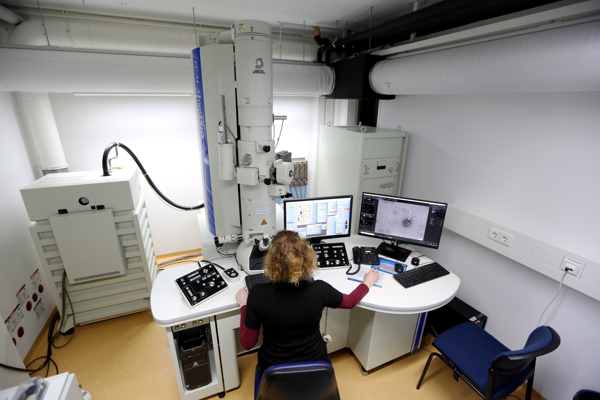
The TEM is a microscope that allows us to take images of biological samples, at a significantly higher resolution and magnification (thousands of times) than common light microscopes. TEM enables our scientists to visualize particularly small structures (Nanometer scale) such as viruses, or bacterial and eukaryotic cells and their substructures (e.g. the membrane or small organelles).
How does a Transmission Electron Microscope work?
During TEM a beam of electron is transmitted through a sample of interest that is adsorbed to a small copper slide (TEM grid). The image is formed when the electrons interact with the material on the grid. Parts of the beam are transmitted depending on how transparent the sample is. Electrons that are emitted from the sample are used to build the image.
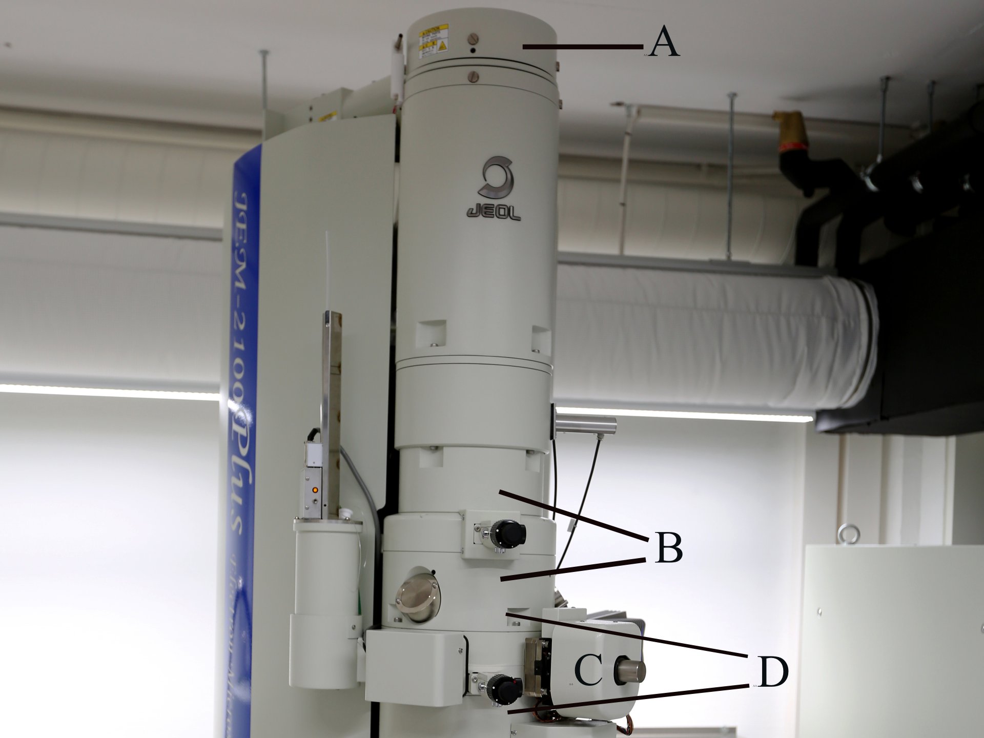
The electron beam is generated in the electron gun at the top of the TEM (A). The gun accelerates the electrons to extremely high speeds using electromagnetic coils and voltages of up to 200kV in our instrument. The beam is then focused into a thin beam by a condenser lens (B) before it hits the sample (C). Emitted electrons are focused into an image by the objective lens (D).
What are we using the Transmission Electron Microscope for?
Viruses infecting ancient microorganisms
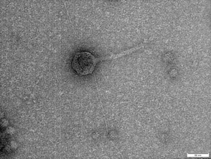
The Archaeal Virology group at the MPI for Marine Microbiology is studying viruses and membrane vesicles of microorganisms that are thought to be very ancient, the Archaea (Ancient Greek meaning: ‘ancient things’). Viruses are very small structure ranging in size from 20-200nm and can only be observed by TEM. The group is isolating and characterizing new viruses, virus-like elements and membrane vesicles of Archaea with the goal to shed light on the origin and evolution of viruses.
Archaeal viruses are very distinct from viruses infecting other organisms such as Bacteria or Eukaryotes (e.g. plants, humans). To characterize these viruses and virus-like elements we need TEM to observe the structure of the virus particles, allowing us to draw conclusion about their classification, their life cycle and their relationships with their host organisms.
Symbiotic relationships under the TEM
The Symbiosis Department studies the biology, ecology and evolution of symbiotic associations between bacteria and marine invertebrates. We investigate these symbioses over a wide spatial range, from the organization of host tissues that house the symbionts down to cellular and subcellular scales of host and symbiont ultrastructure using correlative light and electron microscopy in which fluorescent imaging is combined with high-resolution morphological analyses.
Research questions the Symbiosis Department asks include: How specific are ectosymbiotic bacteria to their hosts, and how do they attach to them? How do endosymbionts enter their hosts, are they extra- or intracellular, and which cellular structures are they associated with? What role do outer membrane vesicles, pili, and other bacterial structures play in interactions of the symbionts with their hosts?
What does EDS mean?
EDS stands for energy-dispersive X-ray spectroscopy. It’s a microanalytical technique used for the chemical characterization of a sample. When the electron beam of TEM hits the sample surface we get an interaction between the electrons of the beam with the atoms in the sample. This interaction produce an X-ray radiation which is specific for every element. This characteristic X-radiation is used to identify and to quantify the chemical elements in the sample.
Technical Details
Instrument
JEOL JEM-2100PLUS
Acceleration voltage: 80, 100, 120, 160, 200kV (Aligned at 80kV, 120kV and 200kV)
Resolution: Point: 0.23nm; Lattice: 0.14nm
Special features and accessories
Speciment holders: High Tilt Room Temperature Retainer (EM-21311 HTR). ±60° tilt
Quick Release Room Temperature Retainer (EM-11610 QR1) ±20° tilt
Cryo-EM holder: Fischione 2550 with tomography capabilities
Camera: Emsis XAROSA 20 Megapixel bottom-mounted CMOS camera
Scanning-Image observation: STEM Detektorsystem (EM-24550YPD) installed
Serial EM (Tomography) installed
EDX detector: Bruker Quantax 200-STEM with XFlash 6, 60mm²
Sample preparation
For sample preparations of suspensions a Leica EM GP2 Automatic Plunge Freezer is available.
For tissue samples a Leica EM ICE High Pressure Freezer, a Leica AFS Freeze substitution unit and two Leica UC7 microtomes are available.
Who uses the Transmission Electron Microscope?
The TEM is mainly used by the Max Planck Research group Archaeal Virology and the Department of Symbiosis. The EDX is used by the Department of Biogeochemistry. However, other researcher at the institute and guest researchers can use the instrument if necessary. Such interdisciplinary cooperation between several working groups results in exciting projects from which all participants can benefit.
Please direct your questions to
Max Planck Research Group Archaeal Virology
MPI for Marine Microbiology
Celsiusstr. 1
D-28359 Bremen
Germany
|
Room: |
2129 |
|
Phone: |

Scientist
MPI for Marine Microbiology
Celsiusstr. 1
D-28359 Bremen
Germany
|
Room: |
3130 |
|
Phone: |


