On Land

The human eye is unable to recognize things smaller than 0.2 mm. At the Max Planck Institute for Marine Microbiology, we study micro-organisms from the environment that are many times smaller – some of them measure only 500–1000 nm. Light microscopes are used to detect these microbes. However, individual structures in the cells can be visualized only with special microscopes.
Moreover, when microbes live together with each other or with animals, everything involves small molecules. From nutrition to communication, proteins, lipids, and the like are almost always involved. We look at these molecules in order to gain insight into the processes both within and between cells.
For this, we use various devices in our laboratories.
Other instruments and techniques
Microscopes
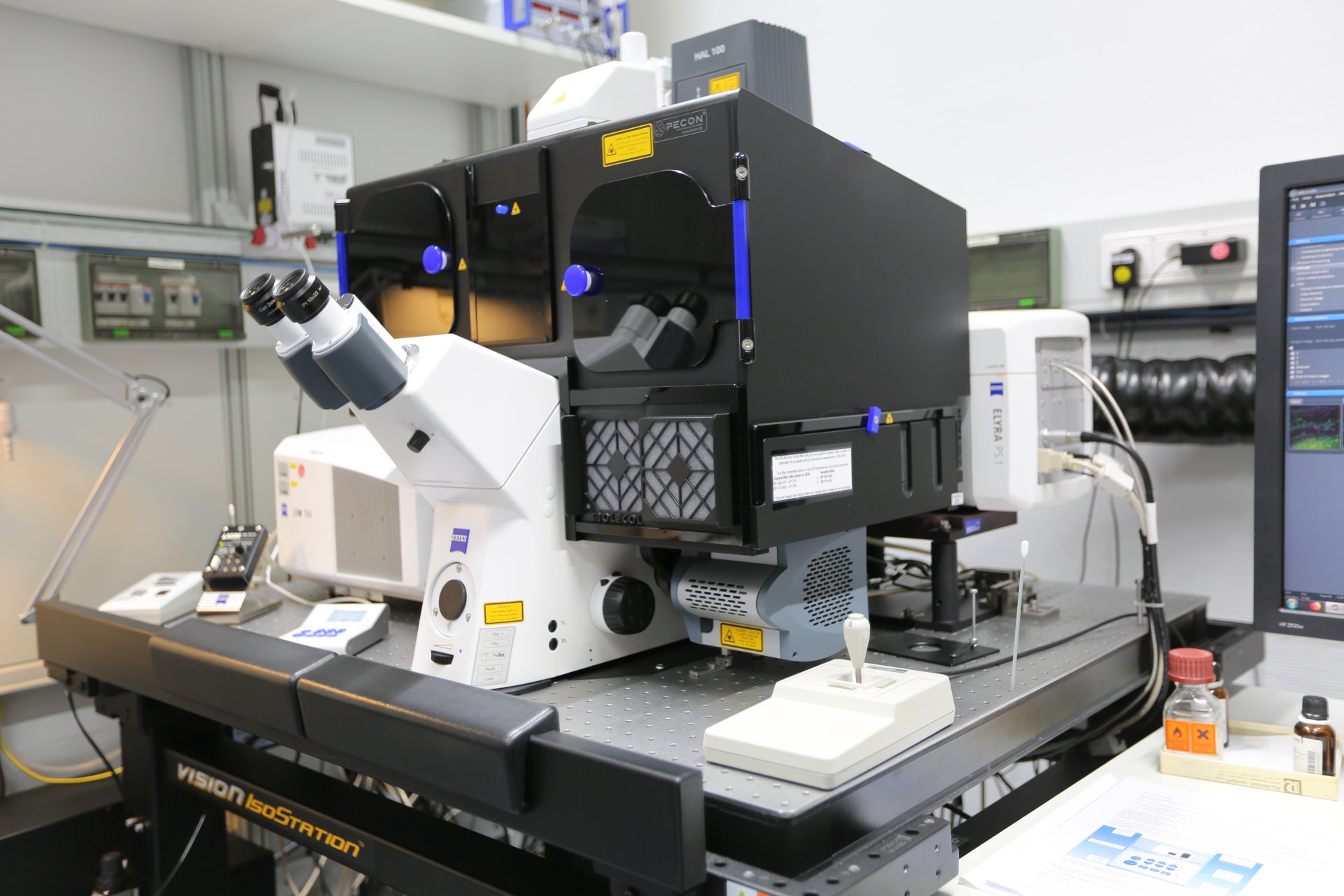
Confocal laser scanning microscope
The confocal laser scanning microscope is our workhorse for detecting specific fluorescence signals. The main task of a confocal microscope is to display as accurate an image of minute objects as possible. It allows a more detailed view of the microbes than a standard light microscope. More…
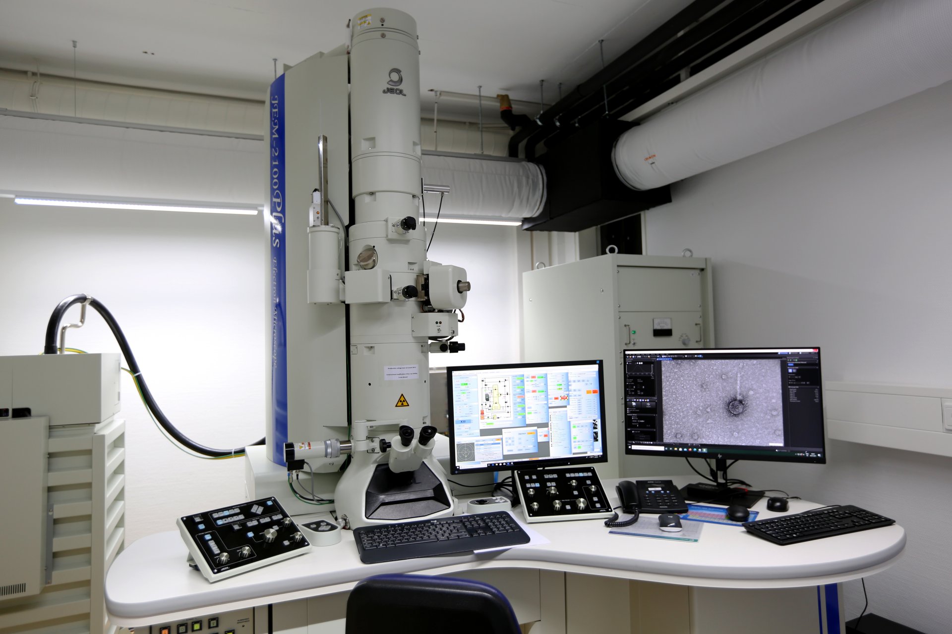
Transmission Electron Microscope (TEM) with EDX detector
The TEM is a microscope that allows us to take images of biological samples, at a significantly higher resolution and magnification than common light microscopes. TEM enables our scientists to visualize particularly small structures at Nanometer scale such as viruses, or bacterial and eukaryotic cells and their substructures like the membrane or small organelles. More...
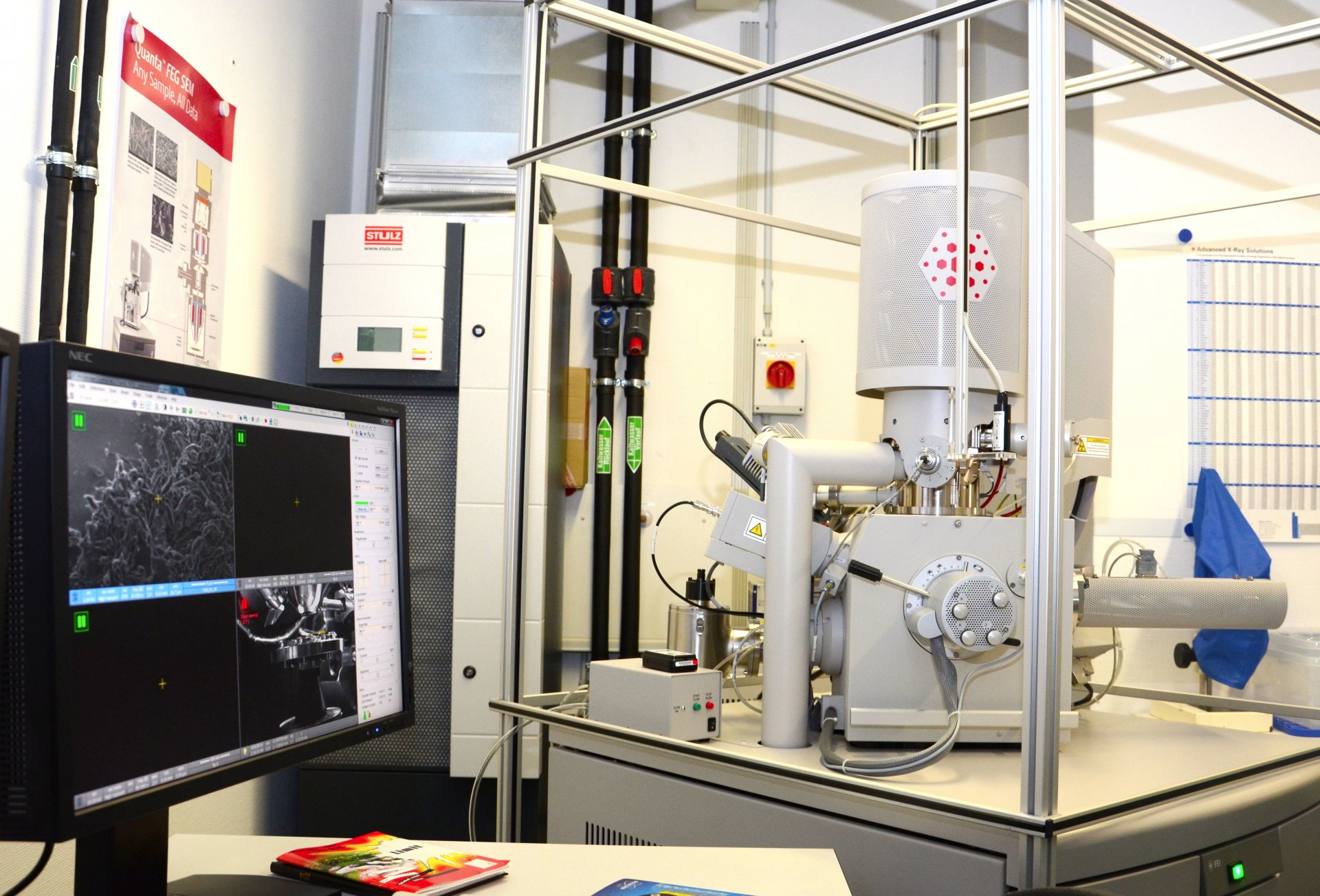
Environmental scanning electron microscope
When examining and imaging very small objects, such as marine microorganisms, one very quickly reaches the limits of optical microscopy. To be able to image these organisms, the application of scanning electron microscopy is the next step. Our scanning electron microscope is equipped with various detectors for image generation and analysis. More...
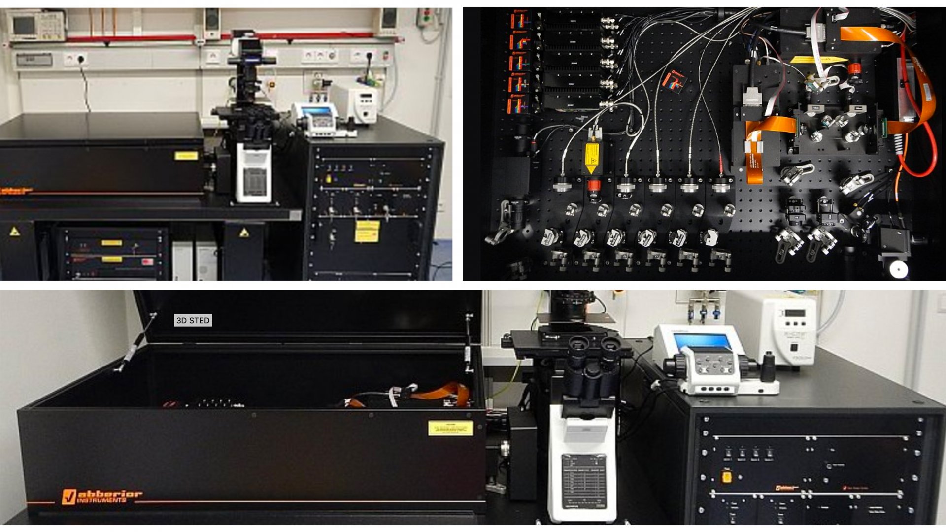
Superresolution microscopy - Stimulated Emission Depletion (STED)
Superresolution microscopy allows us to visualize microstructures below the diffraction limit. Our latest superresolution instrument is based on the Stimulated Emission Depletion (STED) technique. The Abberior Instruments easy3D STED can provide a lateral resolution below 25 nm and a 3D resolution of up to 60 nm. The instrument includes the methodes pulsed-STED, gated-STED, and RESCue STED. It is the first STED microscope with MINFIELD technique on the commercial market.
More information on our device can be found here, at the Department of Molecular Ecology
More information on the function can be found here... (Link to the MPI for Biophysical Chemistry)
Video (Youtube): Stefan Hell explains superresolution and STED
Automated Microscopy and Cell Counting
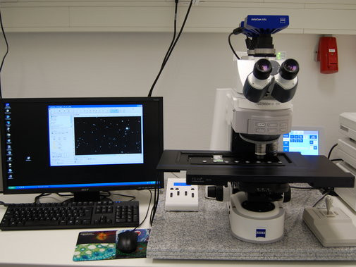
We developed an image acquisition and evaluation system for the automated microscopic evaluation of comparatively simple samples, i.e. the counting of bacterial cells in suspension that are filtered and immobilized on membrane filters. For this we have developed a robust computerized focussing system that combines a proprietary autofocus with custom texture parameters, feedback loops for image contrast maximation, and decisions on image content during acquisition. The system is capable of excluding inappropriate or badly focussed microscopic fields prior to evaluation without operator interaction with high reliability. More...
Mass spectrometers
Nanoscale Secondary Ion Mass Spectrometer (NanoSIMS)
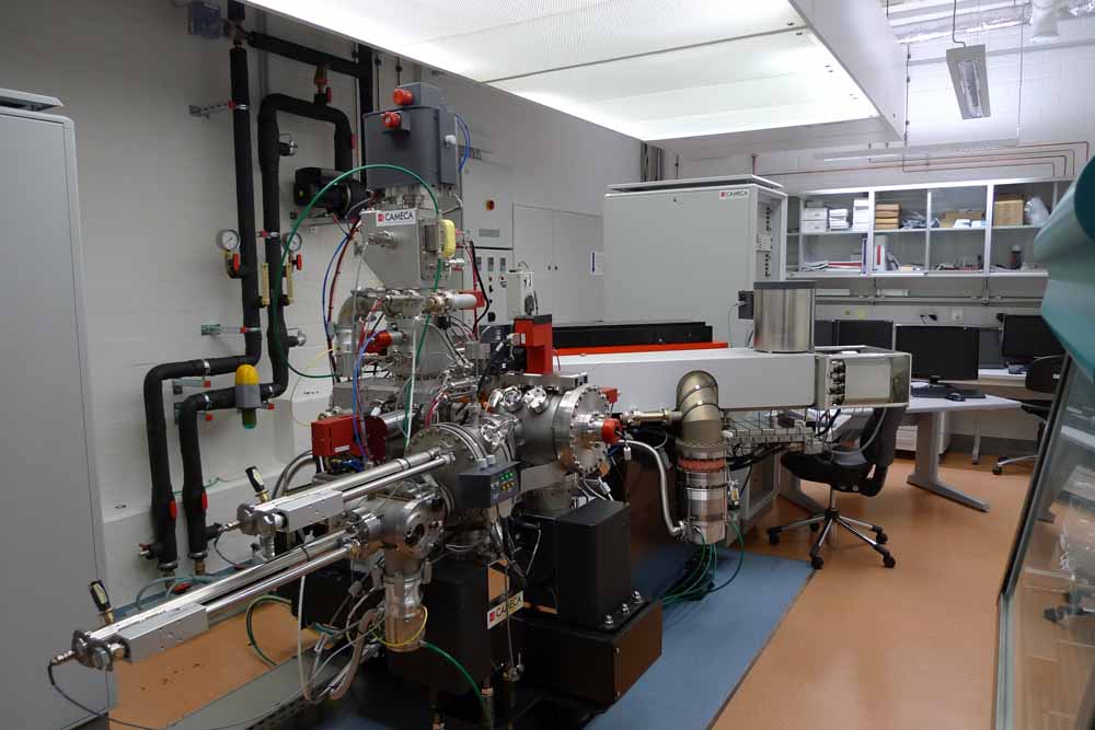
It is big, silver, and looks rather complicated. But it works wonders. With its help, we can watch micro-organisms at work. The NanoSIMS is a mass spectrometer with special optics that enable impressive spatial resolution. We can thus observe things that are only about 50 nm in size. We use it to study structures and processes inside bacterial cells. More...
MALDI imaging mass spectrometry
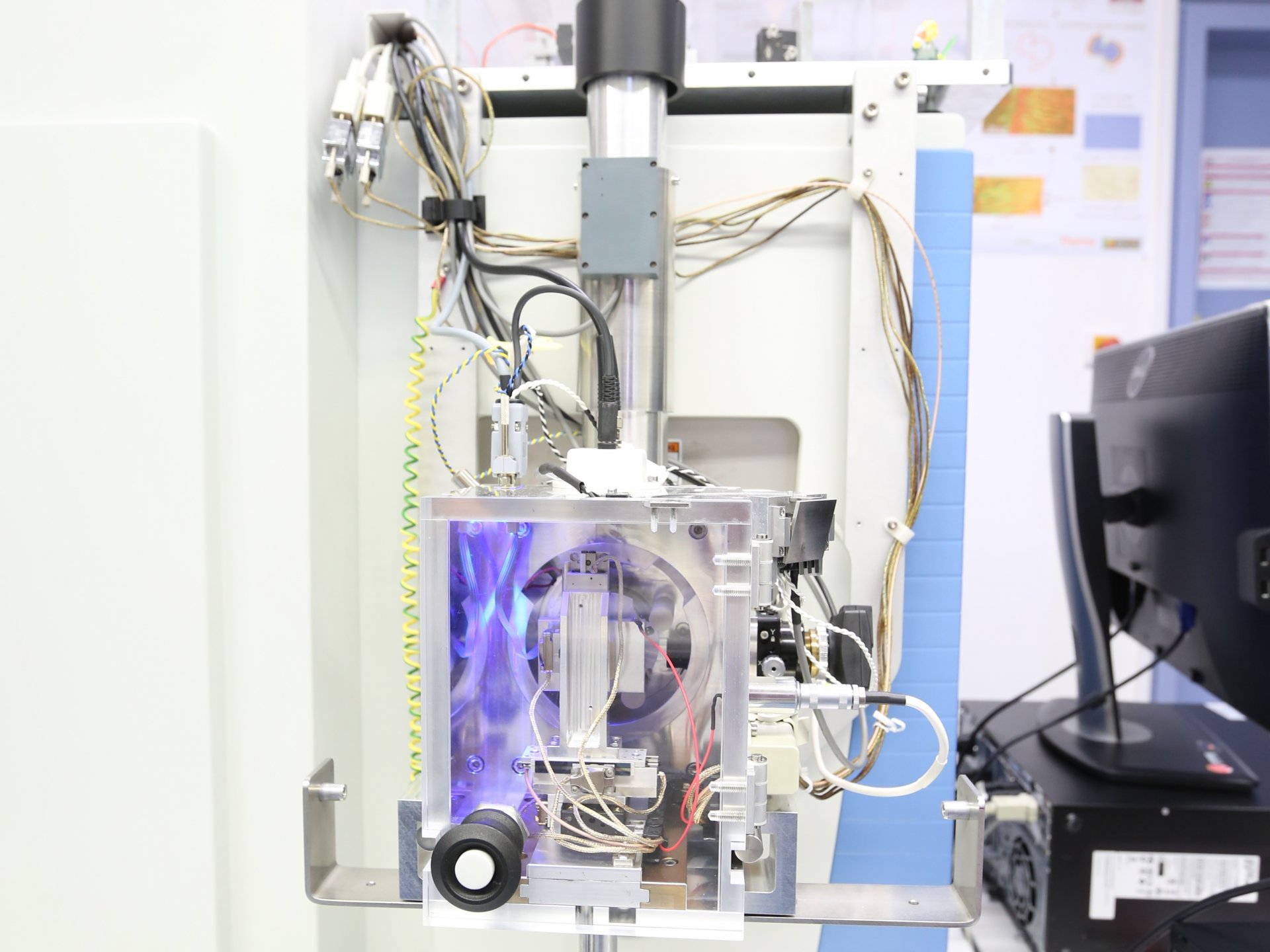
MALDI imaging mass spectrometry is an imaging method for analysing chemical compounds and their spatial distribution in a sample. It can be used to identify where certain compounds are located in a sample. Scientists can use this information to draw conclusions about how the metabolism of the mussel works. More…
High performance liquid chromatograph with mass spectrometry (HPLC-MS)
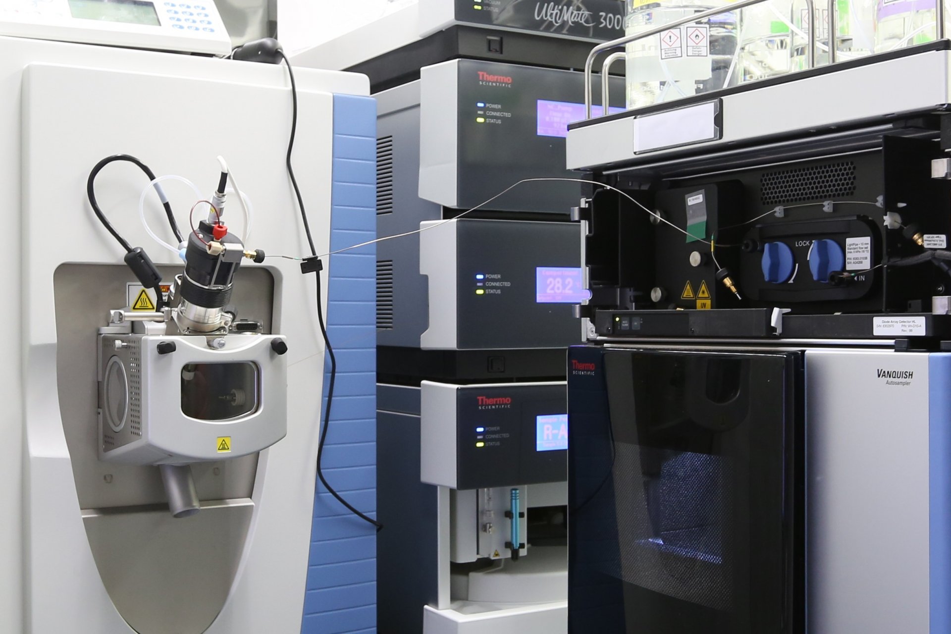
A high-performance liquid chromatograph, coupled to a mass spectrometer, is among the most sensitive instruments available to science. The HPLC-MS allows it to separate mixtures of substances into their components and to measure the molecular masses of these components very precisely. More...
Gas chromatograph with mass spectrometry (GC-MS)
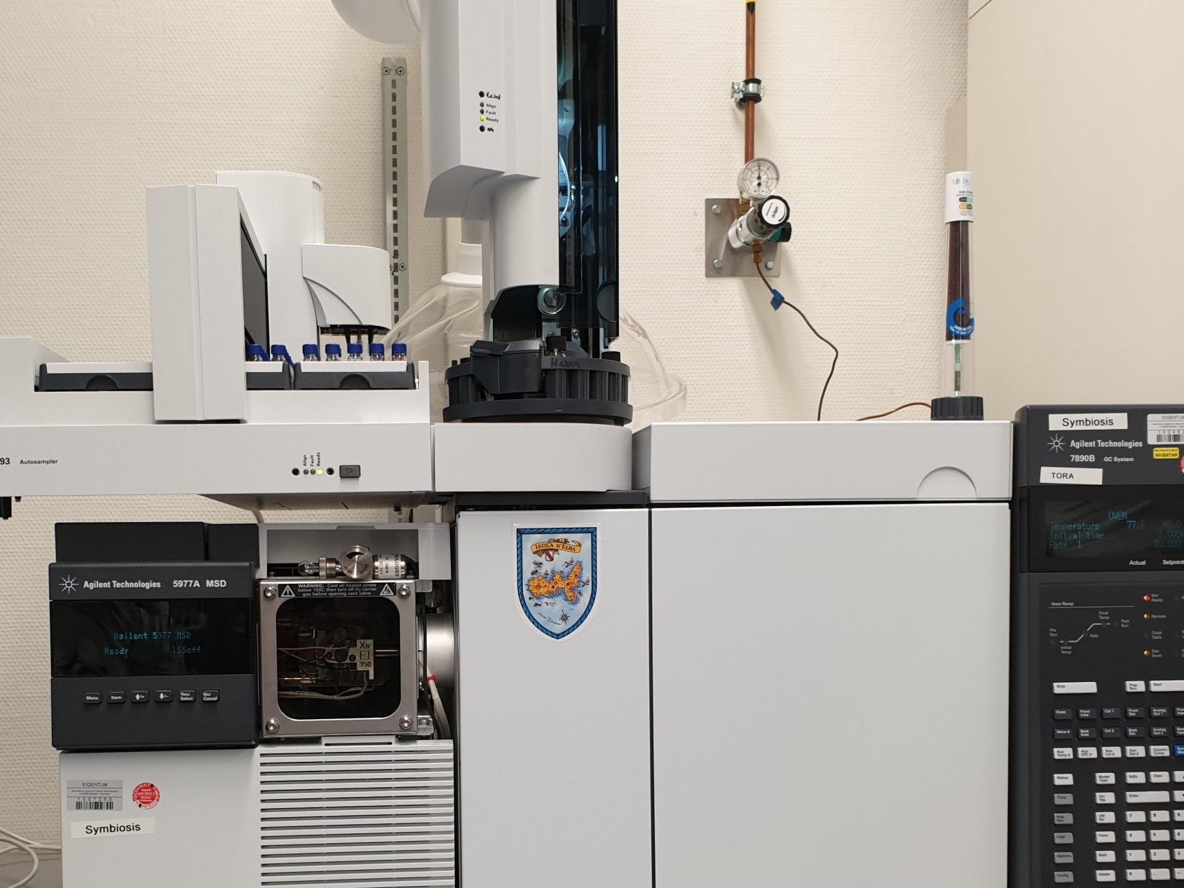
A gas chromatograph can be used to analyze volatile compounds. The connected mass spectrometer serves to determine which substances are contained in the sample and how much of each substance is present.
Ultrahigh-resolution mass spectrometry
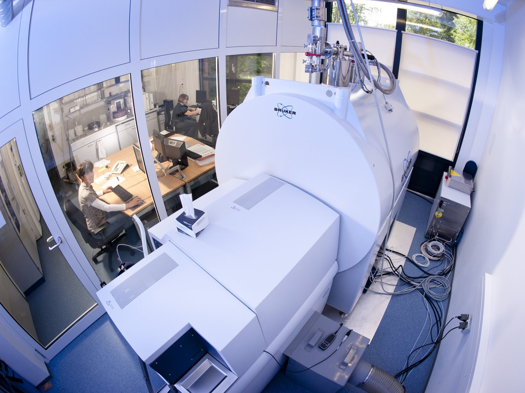
The Research Group for Marine Geochemistry has a Fourier-Transform Ion Cyclotron Resonance Mass Spectrometer (FT-ICR-MS) at its disposal, which offers ultrahigh-resolution mass spectrometry for our research. It is the most powerful technique to molecularly characterize complex organic mixtures, such as DOM, whose composition is still largely unknown. We can determine the mass of an individual molecule with a precision of one ten-thousandth of a Dalton, which is less than the mass of an electron. This level of precision is required to identify individual molecules in seawater.
This special mass spectrometer is located at the Institute for Chemistry and Biology of the Marine Environment of the University Oldenburg (ICBM) and is used by the Research Group Marine Geochemistry, which is a Bridge Group between the ICBM and the Max Planck Institute for Marine Microbiology. The ICBM is the only institution in the marine sciences worldwide that hosts such unique mass spectrometer.
More information is available at the departments page of the Group Marine Geochemistry and at the homepage of the ICBM.
Other instruments and techniques
Chemostat
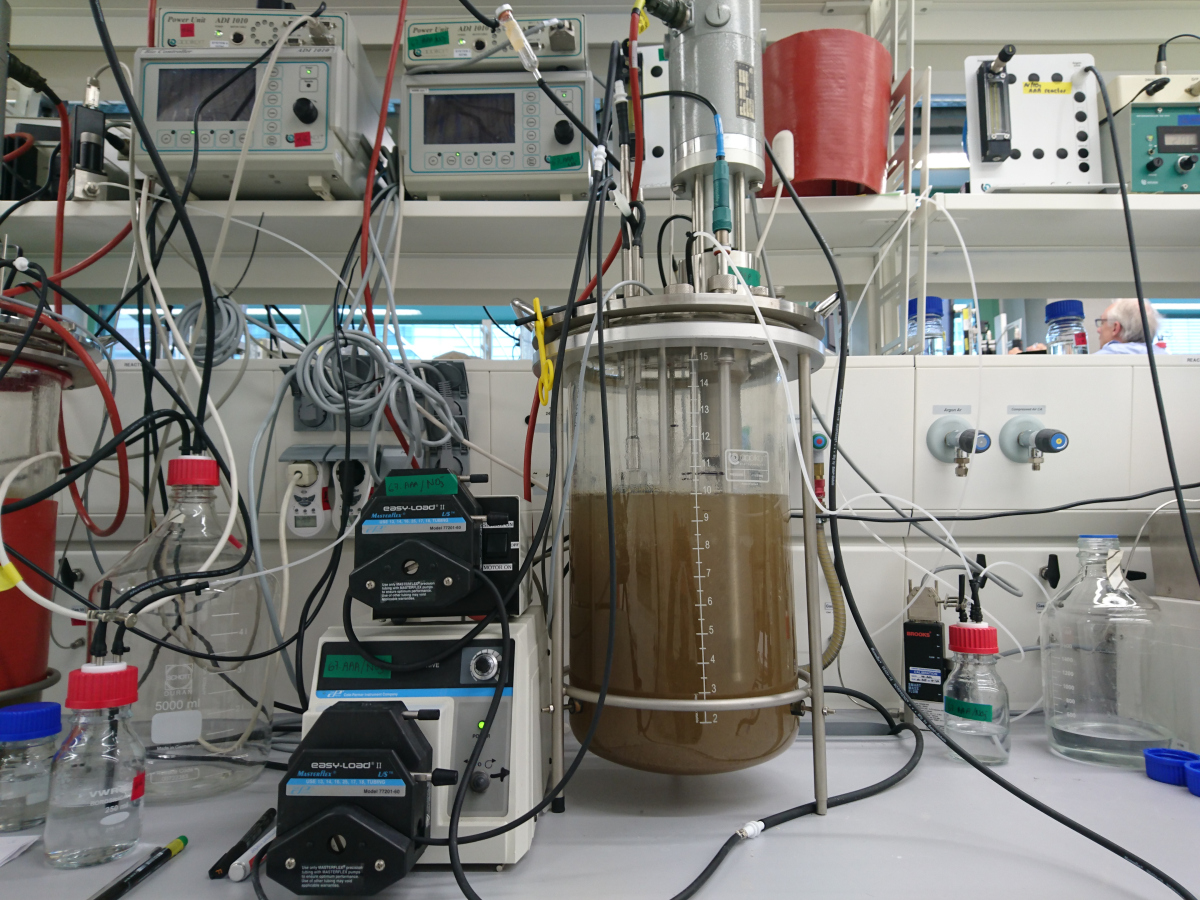
A chemostat is a cultivation bioreactor, which closely mimic the environments that microorganisms live in – in the laboratory, under controlled conditions. That allows us to culture environmentally-relevant microorganisms to study their physiological and biochemical properties in molecular detail. More...
Flow Cytometry
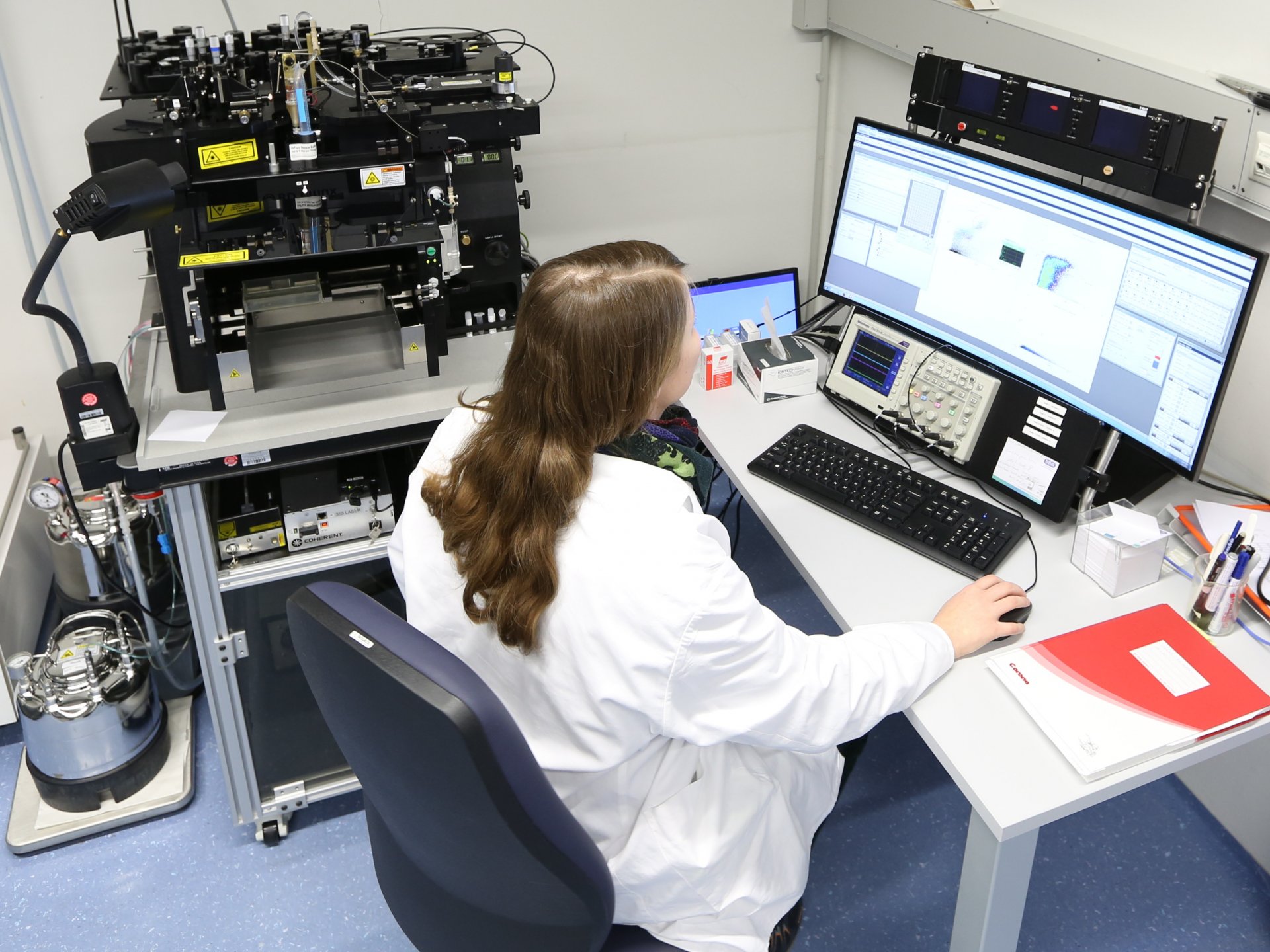
Generally, flow cytometry is the measurement of single cells in a water jet. At the Max Planck Institute for Marine Microbiology, we use flow cytometry routinely to quantify cell numbers picoplankton samples and cell cultures. Further, cells meeting predefined properties can be sorted with high speed in high purity for further molecular biological analysis. More...
Raman spectrometer and atomic force microscope
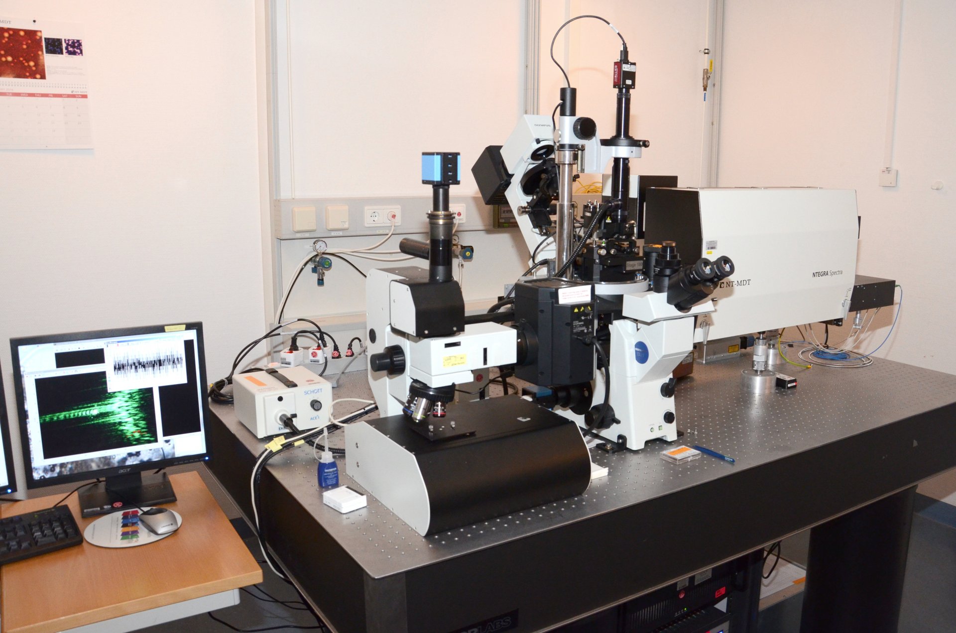
The Raman spectrometer is used to investigate material properties. The great advantage of the device is that it allows non-destructive and non-contact measurement. Furthermore, only very small amounts of a biological sample are needed. More...
Any solid material in air or liquids can be examined using atomic force microscopy. This is a great advantage for biological samples because they will not dry out and thus retain their shape. Information on the surface structure can be determined in the nanometre range. More...
Microsensors
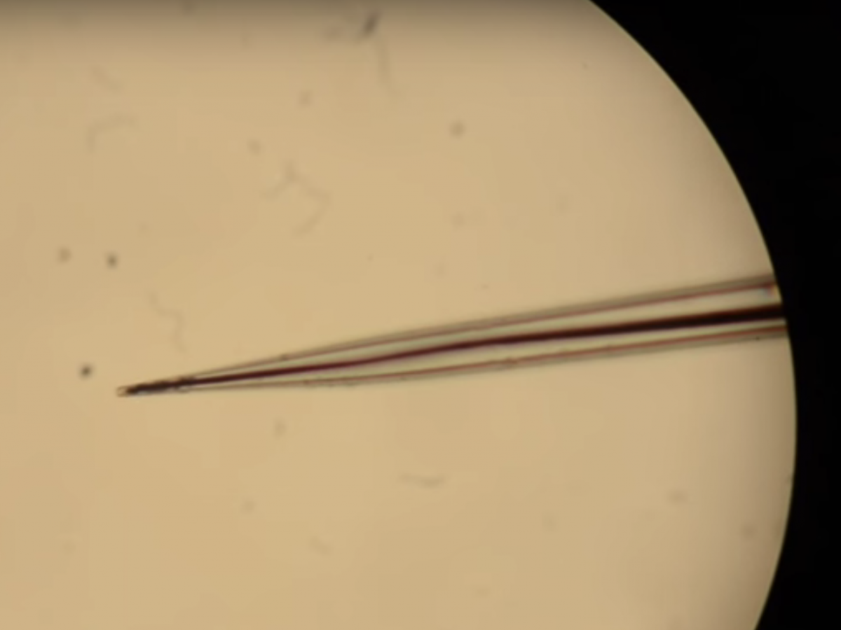
The Microsensor Group studes microbial ecology in a wide range of environments: seep systems, deep-sea and coastal sediments, coral reefs, anoxic lakes, microbial mats, animal-associated microbes and more. The research is highly diverse, encompassing themes of photosynthesis, nitrogen and sulfur cycling, calcification, habitat mapping, ecosystem productivity and cell physiology. Most themes involve the study of the functioning of intact microbial communities with non-invasive methods that allow as direct an observation as possible. To this end, we develop, construct and use microsensors for laboratory and in-situ measurements. These tiny sensors, typically made of extruded glass, have tip sizes in the order of 5-50 μm and can rapidly measure chemical fluxes caused by cells.These microsensors are also applied towards measuring ecosystem fluxes with the eddy covariance technique.
